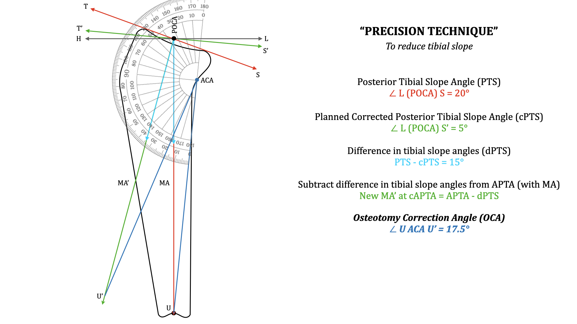
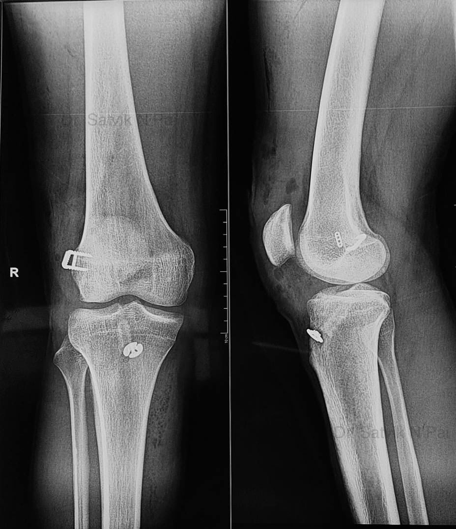
A novel “Precision technique” for preoperative planning of tibial slope correction osteotomy. High Tibial Osteotomy and Tibial Slope Correction Osteotomy requires precise preoperative planning. Tibial slope measurement and a slope correction osteotomy therefore requires planning without errors. Currently utilised preoperative planning techniques for slope correction are not very accurate. These can lead to errors in surgical correction and therefore impact the results. This novel “Precision technique” of planning overcomes errors in planning of the current traditional technique. It gives us the precise osteotomy correction angle by a simple measuring technique. The open access article which is attached highlights and gives all details of this novel technique. It can help one plan precisely, for accurate corrections to get the desired results. Link to the article - https://lnkd.in/gPuwrymD
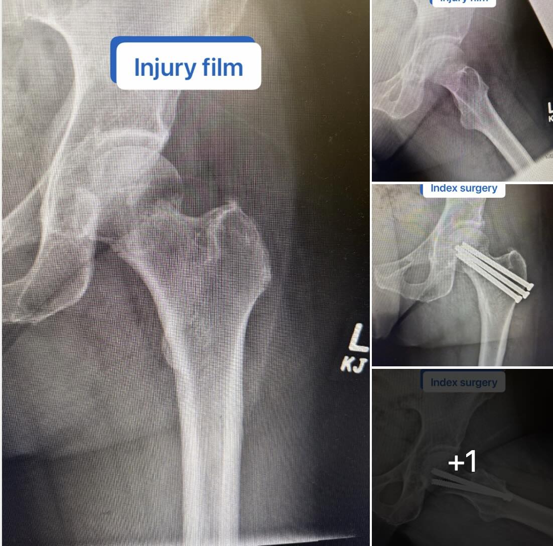
Lateral Hip Pain After Femoral Neck Fixation: What’s the Next Step? A patient presented after successful healing of an impacted femoral neck fracture treated by outside surgeon 5 years ago with closed reduction and percutaneous pinning. The fracture is healed—but now they’re dealing with persistent lateral hip pain. On exam: focal tenderness over the lateral screw heads. This is a scenario many of us see. Read more
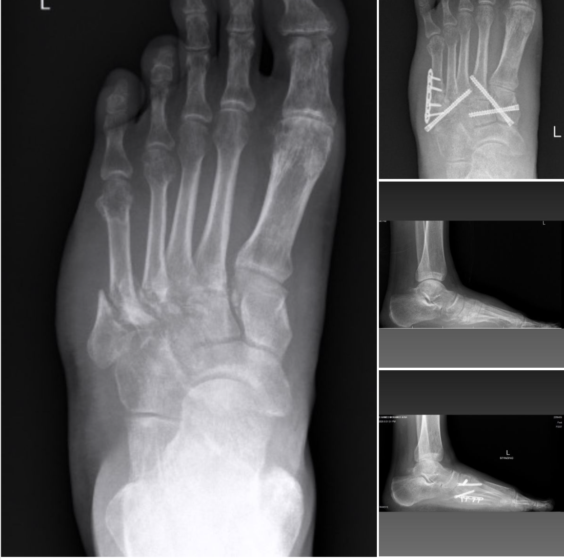
❓For Lisfranc injury in younger patients, which fixation method do you prefer, transarticular screws with early removal, or a bridging plate construct?
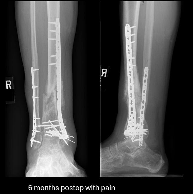
This young patient was 6 months out from my treatment of an open pilon fracture, presenting with pain and swelling. The soft tissues were in good condition. There were no signs of infection and labs were normal. Images showed callus but also loss of coronal alignment, nonunion, and broken implants. The injury and immediate postop films are available for review. Step 1: Is this an extra-articular or intra-articular problem? This one has a healed joint and is an extra-articular issue. Step 2: Mechanical or biologic problem? Primarily a mechanical problem. - Uni-columnar bridge plate likely insufficient to last for the entire duration of healing. - Progressive loss of coronal alignment worsened the environment The biology was affected by initial injury but presence of callus and hypertrophic nonunion suggests it is not the dominant issue Treatment strategy: - Remove failed implants - Restore alignment with a medially based frame Stabilize with medullary nail Reaming stimulated the biology, but the key intervention is mechanical optimization This led to reliable healing in this case. In my experience, medullary nailing with realignment is an excellent option for hypertrophic extra-articular pilon nonunion. What are your thoughts on this problem? Do you like medullary nailing? Any other good treatment options?
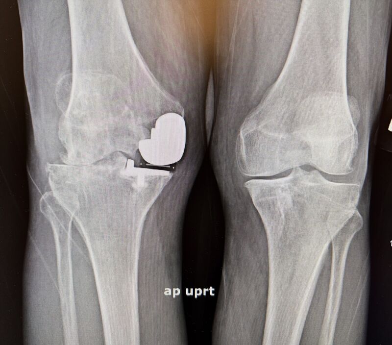
A case that highlights one of the most important principles in knee arthroplasty: patient selection matters. I recently treated a 60-year-old male who presented one year after a medial unicompartmental (partial) knee replacement performed elsewhere. He continued to have significant pain and functional limitation. On evaluation, he had “progressive” arthritis in the lateral and patellofemoral compartments—ultimately requiring conversion to a total knee arthroplasty. I suspect he had significant changes prior to his partial knee arthroplasty, but he was unable to obtain preoperative images. Partial knee replacement can be an excellent operation: Faster recover, More normal knee kinematics, Preservation of bone and ligaments But it is not forgiving when indications are stretched. The key principle: Partial knee replacement is best reserved for patients with truly isolated unicompartmental disease. When arthritis is already present—or likely to progress—in other compartments, the risk of early failure and revision increases significantly. In this case, the patient did well after conversion to a total knee, but it underscores an important point: The success of a partial knee begins before the incision—with proper patient selection. As surgeons, the decision is not just what we can do, but what we should do to give patients the best long-term outcome. Click here to view the original post
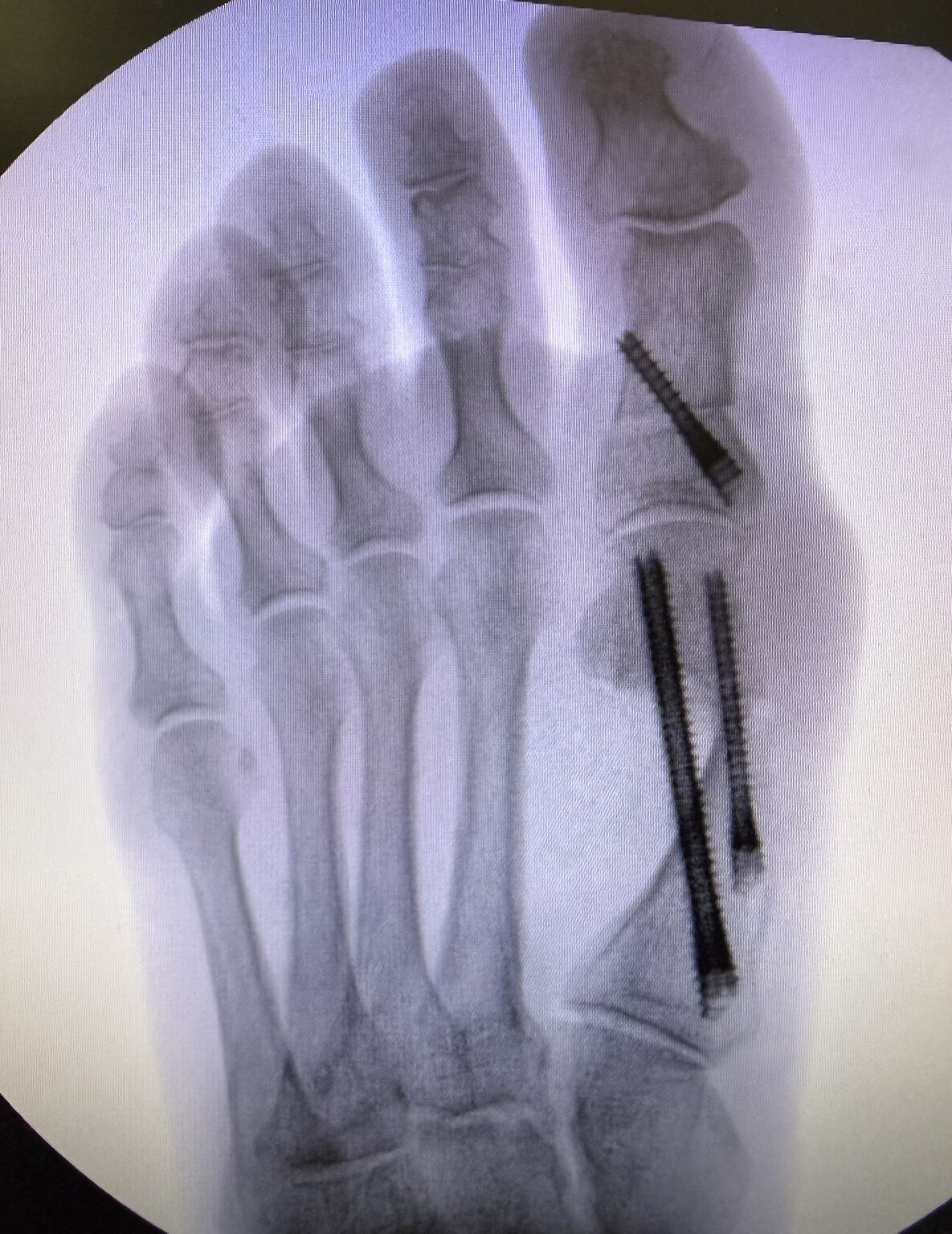
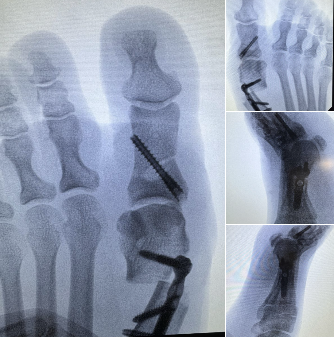
Would you jump from a plane with only one parachute & no reserve parachute?! Not me. Similarly, I’m traditionally a 2-screw fixation surgeon for my first metatarsal during MIS bunion surgery. Maybe 1 screw could get the job done in certain situations & you could get lucky, but why take the risk & have to deal with potential complications if that 1 screw fails? Steps made simple: 1. Place reference wire at osteotomy site. 2. Targeter places wires in perfect position. 3. Make transverse osteotomy. 4. Employ “crow-bar” for 3D deformity correction. 5. Advance wires & place fixation. Do you use a reserve parachute? Click here to view the original post
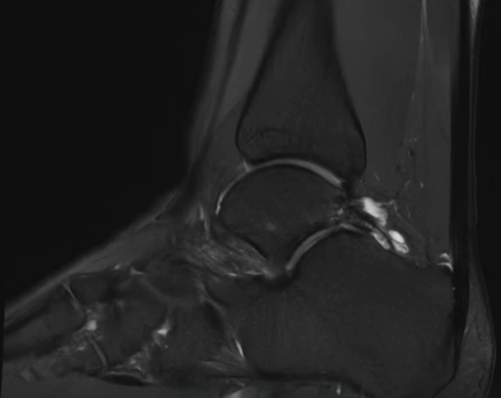
Posterior ankle pain in athletes isn’t always “just impingement.” Often it’s a combined os trigonum + FHL pathology problem. Takeaway from Arthrex TPC Meeting Recent patient with persistent posterior ankle pain—worse with push-off and plantarflexion. Workup: ▪️ Os trigonum on imaging ▪️ Pain with resisted hallux flexion → FHL involvement ▪️ Failed nonoperative management Surgical Decision: Arthroscopic vs Open Arthroscopic approach ✔ Less soft tissue disruption ✔ Faster recovery ✔ Direct visualization of posterior ankle Open approach ✔ Useful for large ossicles or abnormal anatomy ✔ Better for extensive scarring or tenosynovitis ✔ More direct control of the FHL tendon Critical Point If you remove the os trigonum and ignore the FHL… you will miss the primary pain generator in some patients. In this case: ▪️ Posterior ankle arthroscopy ▪️ Os trigonum excision ▪️ FHL tenolysis + release Let’s Talk Technique (for those doing open) Curious how others are approaching this: Medial vs lateral approach? Medial: more direct access to FHL, but tighter working space Lateral: more familiar for os trigonum, but indirect access to FHL Takeaway Posterior ankle impingement is often a two-structure problem Bone + tendon If you’re not evaluating both… you’re not done. Click here to view the original post
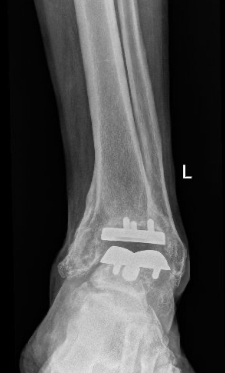
How are you managing failed total ankle replacement? This patient underwent an ankle replacement 5 years ago at an outside hospital. Patient has been in pain and walking on the side of their foot. Slowly getting worse. Clinical Varus. Radiohraphic varus. Infinity tibia and Inbone talus What’s next? - Conversion to fusion - Revision to a replacement - Staged revision Patient underwent removal of implants then revision to Inbone tibia and Invision talus. Frozen & GS negative. Foot deformity needed to be corrected as well. Dwyer and lateral displacement calcaneal osteotomy with base of 1st DF Osteotomy Lateral ligaments stretched out. Modified Brostrum was needed too What would you have done differently? Click here to view the original post
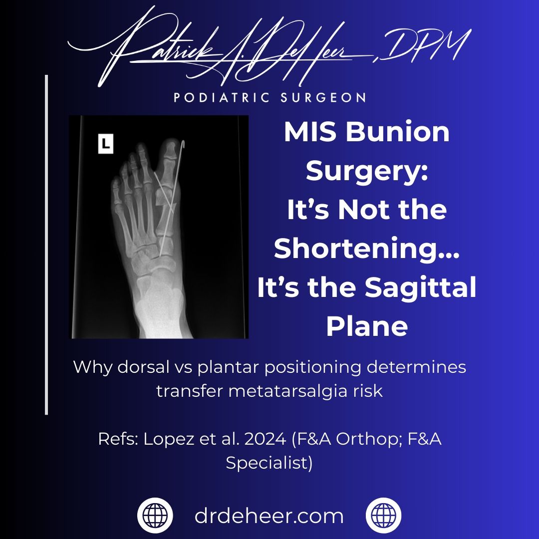
One of the biggest knocks on MIS bunion surgery is transfer metatarsalgia. The assumption has always been that the burr causes shortening and that shortening leads to lateral overload. This study challenges that directly. A 2 mm shortening alone did not increase lateral forefoot pressure. What did? Dorsal displacement of the first metatarsal head. That is the key. If you dorsiflex the capital fragment, you unload the first ray and shift pressure laterally. That is the mechanism for transfer metatarsalgia. If anything, slight plantar positioning restores medial loading. This is not a procedure problem. It is a technique problem. MIS bunion surgery requires precise control of the sagittal plane. That is where the learning curve lives. The takeaway is simple: Respect the first ray. Control the sagittal plane. Do not dorsally elevate the head. That is how you avoid transfer metatarsalgia. Click here to view the original post
Lateral Extra-Articular Tenodesis (LET) with ACL Reconstruction – Are You Adding It? It’s interesting how things can change with time in surgery. LET was actually being performed even before arthroscopic ACL reconstruction became so prevalent. Then arthroscopy took over, and Arthroscopic ACL reconstruction became gold standard. It still is, but LET has definitely made a comeback in the last few years, as an augment atleast. The rotational stability that an LET provides is theoretically and clinically proven beyond doubt. I wanted to specifically talk about two aspects of LET that is evolving — the indications and the method of fixation. First, indications. Considering LET has an added advantage, do you add it to every ACL reconstruction? From not being used at all, we’ve come to stage where atleast some indications we agree on definitely could do with that added rotational stability. We had an international consensus statement published last year that clarified some of it atleast (shared in the comments)- Grade 3 pivot shift, knee hyperextension, revision ACLR, Paediatric ACL’s. I definitely agree with all the indications, but I’ve now started using it in even more. I’ve also been recommending it to all my patients who play any sport, whether is professionally/recreationally, no matter which sport. Cause I don’t necessarily fully agree with the ‘pivoting sports’ indications cause almost any sport with jumping involved can always lead to pivoting on landing. In fact even golf involves pivoting of the knee. Second, fixation of the LET. Interference screw? Anchor? Bone Staple? Soft tissue suturing? I prefer to fix the LET with a bone staple. The time factor is not too significant, but for me the big advantage is not having to worry about femoral tunnel convergence. Sharing an ACL-R + LET done for a professional Kabaddi player. What are your indications to add an LET in a primary ACLR? And how are you fixing it?
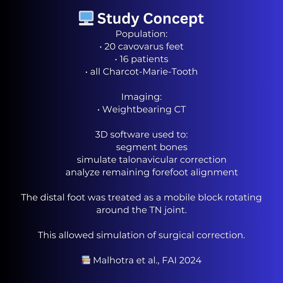
Cavovarus reconstruction is often taught as a series of procedures — calcaneal osteotomy, tendon transfers, first metatarsal dorsiflexion osteotomy, and occasionally midfoot procedures. But it may be more useful to think about the problem from a biomechanical perspective first. The goal of surgery is to restore hindfoot alignment and reduce the talonavicular joint, which re-establishes the normal relationship between the rearfoot and midfoot. Once that occurs, the true residual forefoot deformity becomes apparent. This study used weightbearing CT modeling to simulate talonavicular correction in cavovarus feet and identified four predictable forefoot morphotypes that remain after hindfoot correction. The most common pattern was isolated plantarflexion of the first ray, which explains why a 1st metatarsal dorsiflexion osteotomy is such a common component of cavovarus reconstruction. From a biomechanical perspective, this makes sense. According to Kirby’s rotational equilibrium theory, the foot assumes a position during weightbearing where pronation and supination moments around the subtalar joint axis are balanced. Changes in the relationship between the subtalar axis and the forefoot alter these moment arms and influence how ground reaction forces are distributed across the foot. Once hindfoot alignment is restored, the remaining forefoot deformity represents the mechanism through which the foot is attempting to maintain that equilibrium. Understanding these patterns may help surgeons move beyond generic cavovarus algorithms and toward more precise forefoot balancing strategies that restore a stable tripod and a durable plantigrade foot.
You're seeing a preview. Sign in to see all posts and join the Surgzon community.